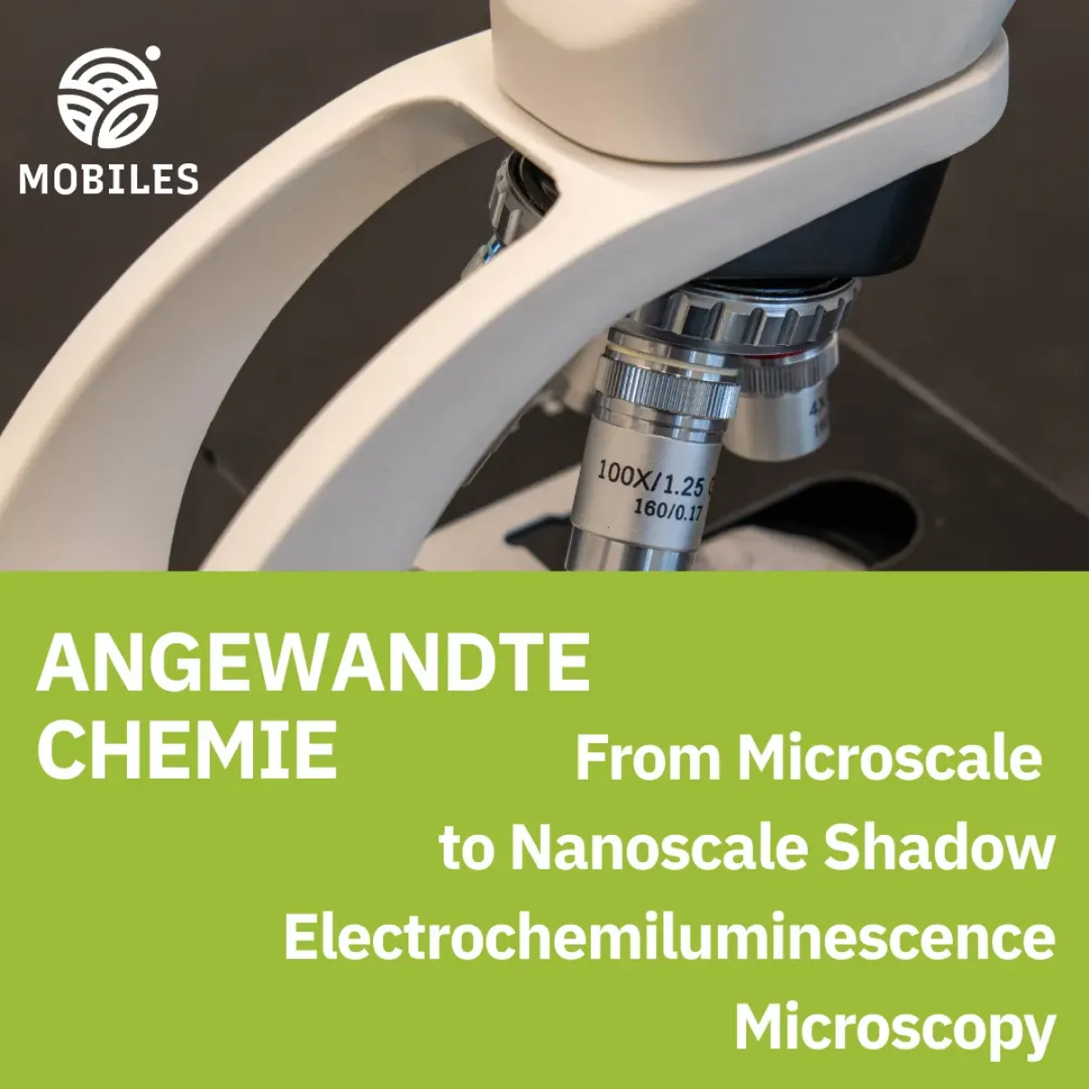
Understanding how to visualize the smallest biological and synthetic particles is essential for advancing diagnostics, materials science, and microbial monitoring. Traditional optical microscopy is limited by diffraction, and even super‑resolution fluorescence techniques face challenges such as photobleaching, background noise, and the need for labels. A recent publication by MOBILES partners demonstrates how shadow electrochemiluminescence (shadow ECL) overcomes these barriers and opens new possibilities for label‑free nanoscale imaging. The study, From Microscale to Nanoscale Shadow Electrochemiluminescence Microscopy establishes the smallest objects ever imaged by shadow ECL and demonstrates its power for complex biological samples, including bacterial spores.
Electrochemiluminescence (ECL) generates light through
electrochemical reactions rather than optical excitation. In shadow ECL, freely
diffusing luminophores create a bright background, while non‑emissive
objects on the electrode surface block electron transfer and reagent diffusion,
producing a dark “shadow.” This
negative‑contrast imaging mode is inherently label‑free and
avoids the phototoxicity and photobleaching associated with fluorescence
microscopy.
The new study systematically explored the detection limits of
shadow ECL. By optimizing electrochemical conditions, optical configurations,
and multi‑frame averaging, the researchers demonstrated that shadow ECL can
reliably detect single nanoparticles down to 100 nm, and in some conditions,
approach 50 nm sensitivity. This represents the smallest insulating particles
imaged by shadow ECL to date.
Two electrode configurations were tested:
Both allowed detection of sub‑micron particles, but ITO offered
superior sensitivity and more consistent linearity between particle size and
shadow contrast. This linear relationship indicates that shadow ECL responds
predictably across scales, a key requirement for quantitative nanoscale
imaging.
Advanced image processing, such as deconvolution and deep‑learning‑based
denoising, further enhanced visibility without
altering the underlying physical signal.
To demonstrate real‑world applicability, the team imaged Bacillus subtilis spores, complex biological structures typically 1–2 µm in size. Shadow ECL successfully captured their morphology and distribution, confirming that the method can handle heterogeneous, irregularly shaped biological samples, not only idealized spherical nanoparticles.
This capability is particularly relevant for microbial monitoring, where spores and other resilient structures play a critical role in contamination, persistence, and surface colonization.
These findings highlight several important outcomes:
By pushing the sensitivity of shadow ECL into the nanoscale, this work opens new avenues for real‑time, label‑free detection of microscopic and sub‑microscopic entities, strengthening the toolbox available for advanced microbial and material monitoring.
From Microscale to Nanoscale Shadow Electrochemiluminescence Microscopy
Autors: Xiaodan Gou, Hanna Manko, Jasmina Vidic, Laurent Cognet, Jun-Jie Zhu, Neso Sojic
Full publication here

