Biofilms pose a persistent challenge across food production,
healthcare, and industrial environments because they allow bacteria to survive
on surfaces despite cleaning and disinfection. Bacillus cereus, a common
environmental bacterium capable of causing foodborne illness and opportunistic
infections, forms biofilms in which cells are embedded in a protective
extracellular matrix. This structure shields the microbial community from
chemical stress and makes removal extremely difficult. Traditional detection methods
rely on culturing and often miss the early, dynamic stages of biofilm
development, underscoring the need for real‑time, surface‑specific
monitoring tools.
A recent study, In Situ Electrochemical Monitoring of
Bacillus cereus Biofilm Formation, carried out by three MOBILES
partners—INRAE, the University of Belgrade, and the University of
Bordeaux—demonstrates how electrical impedance spectroscopy (EIS) can serve as
a sensitive, label‑free method to track biofilm formation directly on
material surfaces.
As bacteria attach, proliferate, and produce extracellular
polymers, they alter the electrical properties of the surface. EIS captures
these changes, particularly variations in charge transfer resistance, and
translates them into a real‑time picture of biofilm
progression. This makes it possible to detect the earliest stages of
attachment, follow maturation, and identify detachment events long before they
become visible.
For MOBILES, this represents a promising pathway toward deployable, on‑site biofilm monitoring technologies that can support safer food systems and cleaner industrial processes.
The study compared biofilm development on two commonly used conductive materials: gold and indium tin oxide (ITO).
These differences suggest that surface chemistry and material properties strongly influence how B. cereus organizes itself, which has direct implications for designing surfaces that either discourage biofilm formation or allow more effective monitoring.
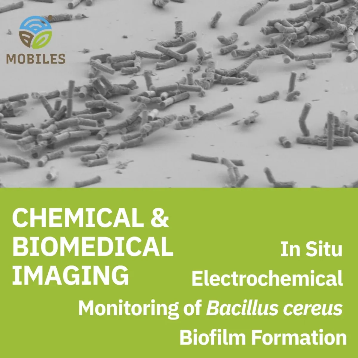
Confocal and scanning electron microscopy confirmed that the electrical changes detected by EIS directly reflect biofilm structure and density. Mature biofilms contained more redox‑active molecules and tighter cell aggregates, which improved conductivity and lowered charge transfer resistance. This alignment between imaging and electrochemistry strengthens the case for EIS as a reliable, real‑time proxy for biofilm architecture.
The study demonstrates that impedance‑based sensors can detect biofilm formation early, non‑destructively, and directly on relevant materials. This has several practical outcomes:
These findings reinforce the value of advanced sensing technologies in developing next‑generation monitoring tools that help prevent microbial contamination and improve environmental and food safety.
In Situ Electrochemical Monitoring of Bacillus cereus Biofilm Formation
Autors: Aleksandar Mijajlovic, Dalibor Stankovic, Milica Sentic, Vlad Costache, Shanshan Wang, Hadi Jbara, Julien Deschamps, Romain Briandet, Neso Sojic, and Jasmina Vidic
Full publication here

